Search Result
Results for "
protein stains
" in MedChemExpress (MCE) Product Catalog:
3
Biochemical Assay Reagents
| Cat. No. |
Product Name |
Target |
Research Areas |
Chemical Structure |
-
- HY-D0914
-
|
FD&C Green No. 3; Food green 3; C.I. 42053
|
Fluorescent Dye
|
Others
|
|
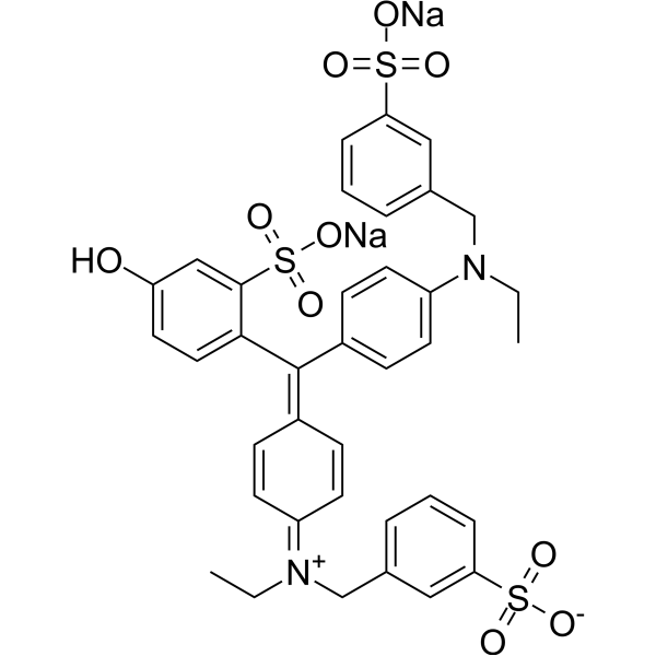
Fast Green FCF is a sea green triarylmethane food dye, with absorption maximum ranging from 622 to 626 nm. Fast Green FCF is widely used as a staining agent like quantitative stain for histones at alkaline pH after acid extraction of DNA, and as a protein stain in electrophoresis. Fast Green FCF is carcinogenic and acts as a presynaptic locus by inhibiting the release of neurotransmitters in the nervous system .
|
-
-
- HY-W127770
-
|
|
Fluorescent Dye
|
Others
|
|
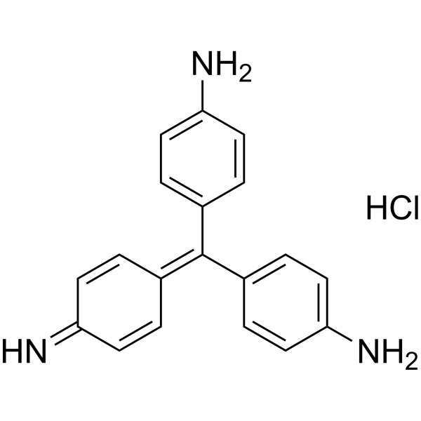
Pararosaniline hydrochloride is a pH-responsive basic dye, as a biological stain to track certain proteins. Pararosaniline hydrochloride has been used in the analysis of SO2 and formaldehyde and staining of bacteria or other organisms .
|
-
-
- HY-D0232
-
|
Brilliant Blue R
|
Fluorescent Dye
|
Others
|
|
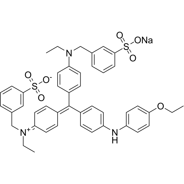
Brilliant blue R250 (Brilliant Blue R), an anionic dye, is the most popular stain to detect proteins resolved in SDS-PAGE gels .
|
-
-
- HY-D1544
-
|
|
Fluorescent Dye
|
Others
|
|
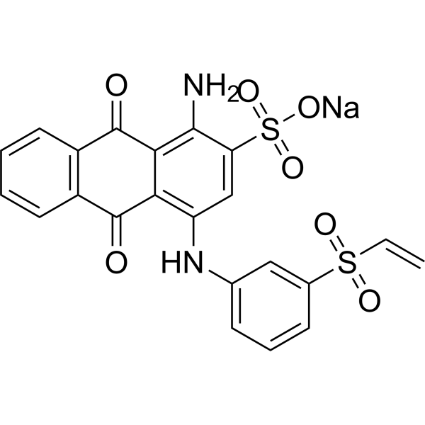
Uniblue A sodium is a reactive protein stain that can be used in the covalent pre-gel staining of the protein (Ex=594 nM) .
|
-
-
- HY-D1674
-
|
|
Fluorescent Dye
|
Others
|
|
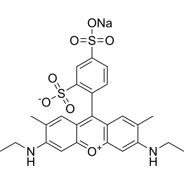
Sulforhodamine G is a fluorescent stain with broad dynamic ranges. Sulforhodamine G can be used for the research of protein stains .
|
-
-
- HY-12489
-
|
Acid Red 112
|
Fluorescent Dye
|
Others
|
|
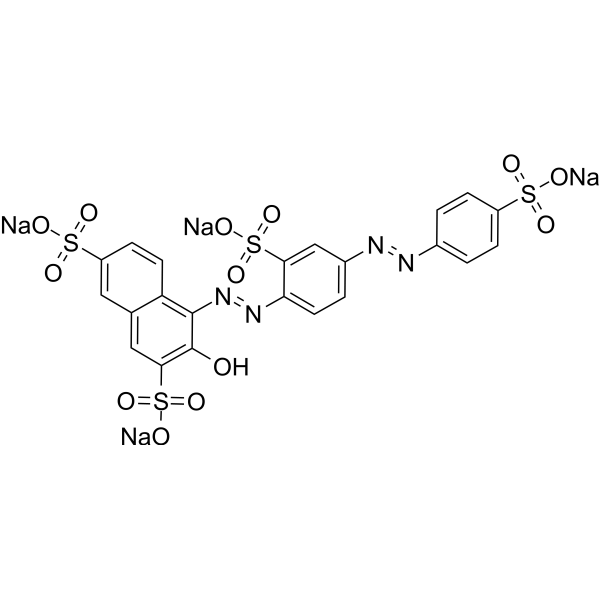
Ponceau S (Acid Red 112) is a non-specific protein dye commonly used as a stain for Western blot. Ponceau S is used in an acidic aqueous solution that is compatible with antibody-antigen binding and dyes the proteins on the membrane red .
|
-
-
- HY-W011831
-
|
3-(Benzyldimethylammonio)propane-1-sulfonate; PDA
|
Biochemical Assay Reagents
|
Others
|
|
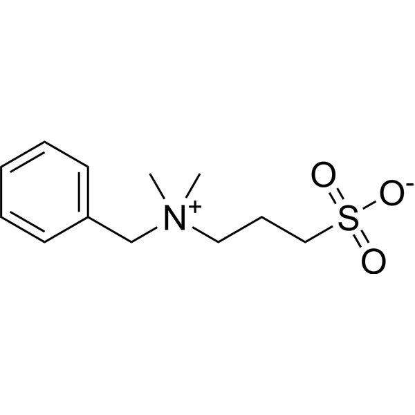
NDSB-256 is a non-stain remover sulfabetaine. NDSB-256 prevents protein aggregation and promotes denaturation of chemically and thermally denatured proteins .
|
-
-
- HY-W133997
-
|
|
Fluorescent Dye
|
Others
|
|
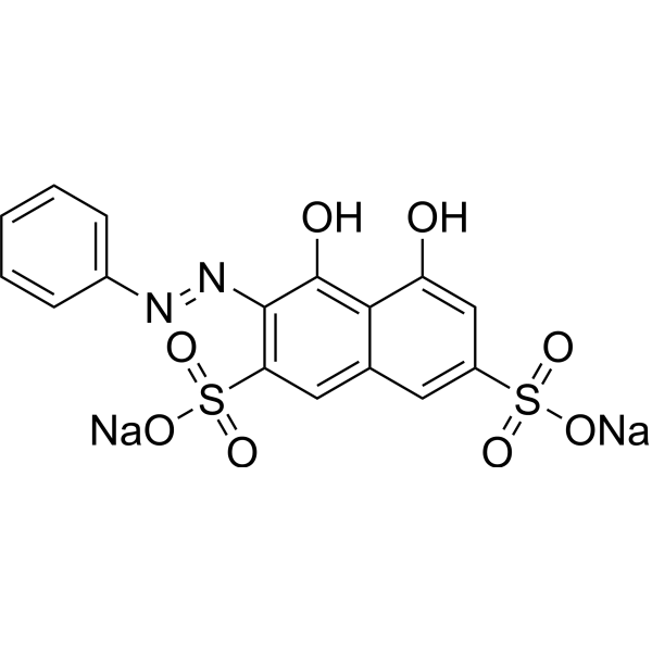
Chromotrope 2R can be used as a chromogenic analytical probe for the quantification of proteins. Basic proteins stained red and the peak wavelength red shifts from 501.6 nm to 567 nm .
|
-
-
- HY-Y0700
-
|
|
Fluorescent Dye
|
Others
|
|
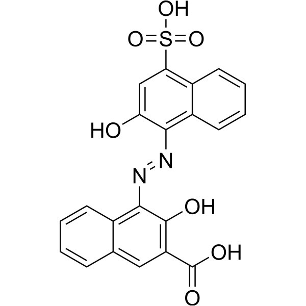
Calconcarboxylic acid, an azo dye, acts as a silver-ion sensitizer to stain protein in SDS-PAGE gels. Calconcarboxylic acid increases silver binding on protein bands or spots by the formation of a silver-dye complex and also increases the reducing power o
|
-
-
- HY-D1475
-
|
|
Fluorescent Dye
|
Others
|
|
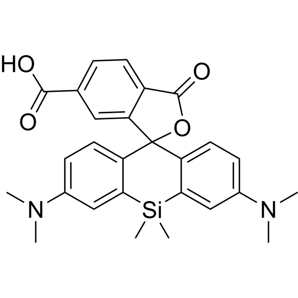
SIR-6-COOH is a fluorescent dye. SIR-6-COOH can be used for staining proteins in live-cell STED imaging
|
-
-
- HY-D1615
-
|
|
Fluorescent Dye
|
Others
|
|
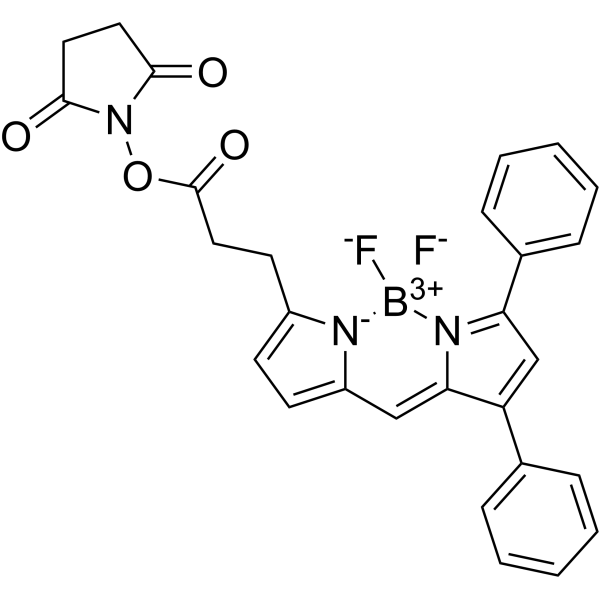
BODIPY 530/550 NHS ester can be used for the stain of protein. BODIPY 530/550 NHS ester can be used for fluorescence OIM (oblique illumination microscopic) image .
|
-
-
- HY-D0987
-
|
|
Calmodulin
|
Others
|
|
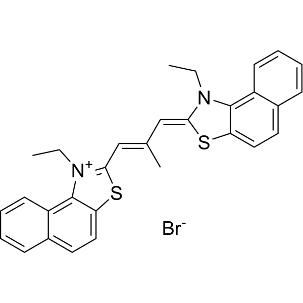
Stains-All, a cationic carbocyanine dye, is a convenient probe to study the structural features of the individual calcium-binding sites of calmodulin (CaM) and related calcium-binding proteins (CaBP) .
|
-
-
- HY-D1543
-
|
|
DNA Stain
|
Infection
|
|
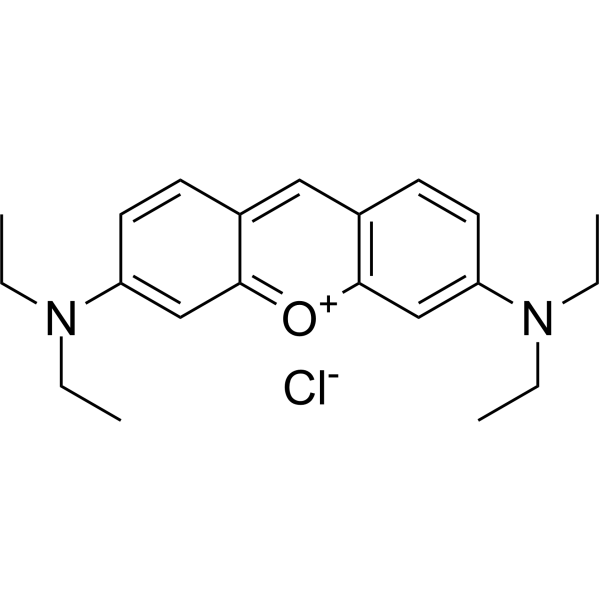
Pyronin B is an organic cationic dye used for the staining of bacteria, mycobacteria and ribonucleic acids. Pyronin B is also used as a small hydrophobic (SH) protein channel inhibitor .
|
-
-
- HY-D1493
-
|
|
PKC
|
Others
|
|
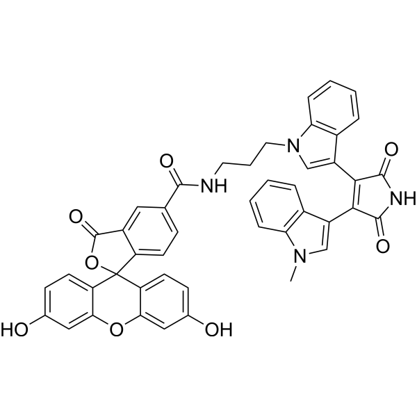
FIM-1 is a fluorescent PKC (protein kinase C) probe that can be used for mitochondrial staining. FIM-1 inhibits PKC and acts as ATP-competitive catalytic site inhibitor .
|
-
-
- HY-D0166A
-
|
|
Biochemical Assay Reagents
|
Others
|
|
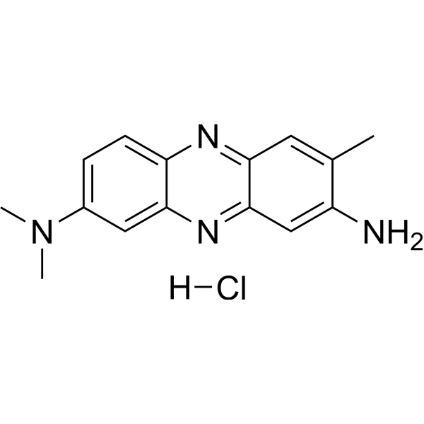
Neutral Red (IND) is an organic dye commonly used in biology and cytology laboratories. It can be used to stain living cells, secreted proteins and other molecular structures, etc., and has a wide range of applications in cell imaging and staining. In addition, Neutral Red (IND) is widely used in industrial fields such as water treatment, food processing and paper manufacturing, for example as an indicator or colorant. Although the compound has no direct medical application, it has important application value in the fields of biology, chemistry and industry.
|
-
-
- HY-12489A
-
|
Acid Red 112, BS
|
Biochemical Assay Reagents
|
Others
|
|
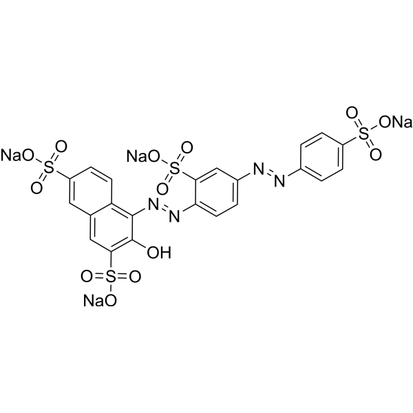
Both Ponceau S and Ponceau BS are synthetic dyes commonly used in biological research. They are commonly used as protein stains to visualize proteins in western blots and other protein detection analyses. Ponceau S is a red dye, while Ponceau BS is a blue shade of the same dye. Both dyes bind to positively charged amino acid residues in proteins for easy visualization. However, Ponceau S is more commonly used due to its higher sensitivity.
|
-
-
- HY-D0014
-
|
|
Biochemical Assay Reagents
|
Others
|
|
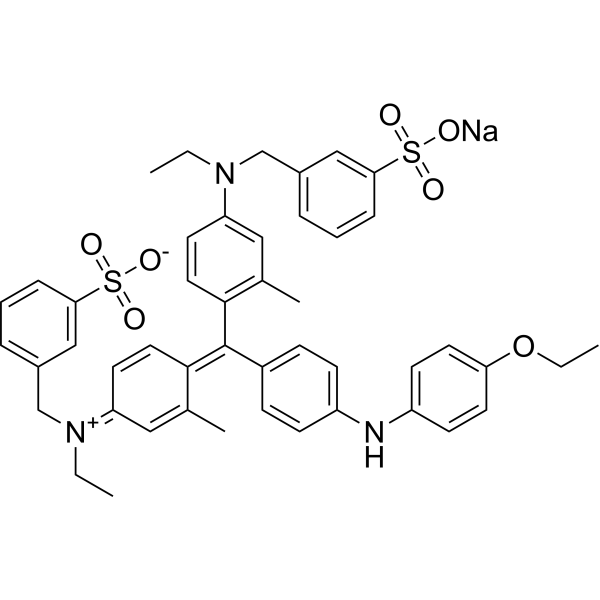
Brilliant Blue G-250 is a dye commonly used for the visualization of proteins separated by SDS-PAGE, offering a simple staining procedure and high quantitation. In the Bradford protein assay, protein concentrations are determined by the absorbance at 595 nm due to the binding of Brilliant Blue G-250 to proteins. Brilliant Blue G-250 is a safe highly selective P2×7R antagonist with promising consequent inactivation of NLRP3 inflammasome .
|
-
-
- HY-W127703
-
|
|
Fluorescent Dye
|
Others
|
|
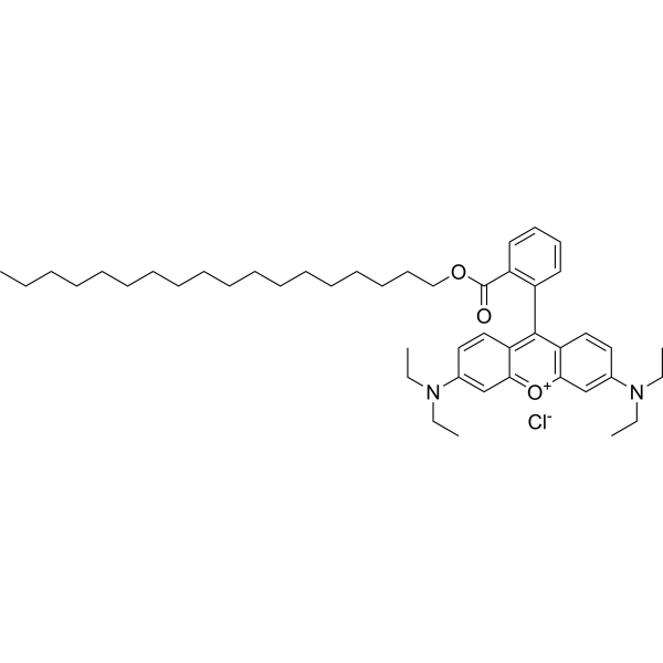
Octadecyl Rhodamine B chloride is a cationic amphiphile that can be used for staining cell membranes. Octadecyl Rhodamine B chloride can be used in numerous studies including electronic energy transfer in organized molecular assemblies, membrane structure, and distances of closest approach between protein domains and membranes .
|
-
-
- HY-135056
-
|
|
Fluorescent Dye
|
Others
|
|
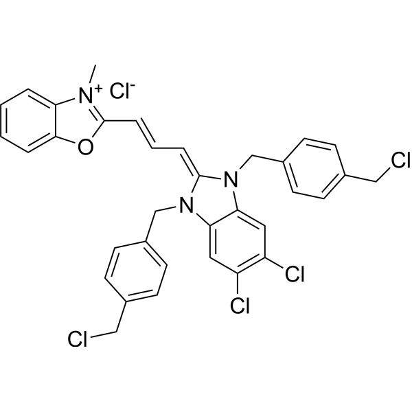
Mito-Tracker Green is a green fluorescent dye that selectively accumulates in the mitochondrial matrix. MitoTracker Green FM covalently binds mitochondrial proteins by reacting with free mercaptan of cysteine residues, allowing staining of mitochondrial membrane potential independent of membrane potential. Excitation/emission wavelength 490/523 nm.
|
-
-
- HY-W133919
-
|
|
Biochemical Assay Reagents
|
Others
|
|
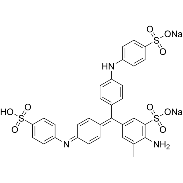
Aniline Blue sodium is a water-soluble dye commonly used as a biological stain for the detection of nucleic acids and proteins in various laboratory procedures such as electrophoresis and microscopy. Aniline Blue sodium has unique chemical properties that allow it to bind to specific cellular components, producing a color change that facilitates their visualization and analysis.
|
-
-
- HY-D1783
-
|
|
Fluorescent Dye
|
Others
|
|
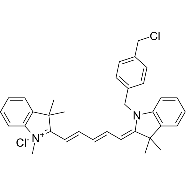
MitoTracker Deep Red FM fluorescent dye that selectively accumulates in the mitochondrial matrix. MitoTracker Deep Red FM covalently binds mitochondrial proteins by reacting with free mercaptan of cysteine residues, allowing staining of mitochondrial membrane potential independent of membrane potential. Excitation/emission wavelength 644/665 nm . Storage: Keep away from light.
|
-
-
- HY-D0938
-
CFDA-SE
Maximum Cited Publications
53 Publications Verification
5(6)-Carboxyfluorescein diacetate succinimidyl ester; 5(6)-CFDA N-succinmidyl ester
|
Fluorescent Dye
|
Others
|
|
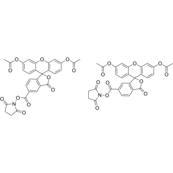
CFDA-SE is a fluorescent dye that can penetrate the cell membrane. It can react with the free amine group in the cytoskeleton protein inside the cell, and finally form a protein complex with fluorescence. After entering the cell, CFDA-SE locates in the cell membrane, cytoplasm and nucleus, and the fluorescence staining is strongest in the nucleus .
CFDA-SE dye can be uniformly inherited by the cells with cell division and proliferation, and its attenuation is proportional to the number of cell divisions. This phenomenon can be detected and analyzed by flow cytometry under the excitation light of 488 nm, and can be used to detect the proliferation of cells .
|
-
-
- HY-138658
-
|
JF526, SE; JF526, NHS
|
Fluorescent Dye
|
Others
|
|
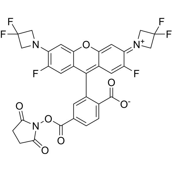
Janelia Fluor 526, SE (JF526,SE) is a fluorogenic yellow fluorescent dye that contains NHS ester group. JF526 is a versatile scaffold for fluorogenic ligands, including labels for genetically encoded self-labeling protein tags and stains for endogenous structures . Janelia Fluor products are licensed under U.S. Pat. Nos. 9,933,417, 10,018,624 and 10,161,932 and other patents from Howard Hughes Medical Institute.
|
-
-
- HY-D0079
-
|
Hydroethidine; PD-MY 003
|
Fluorescent Dye
|
Others
|
|
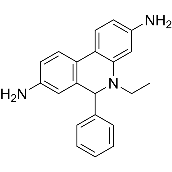
Dihydroethidium, also known as DHE, is a peroxide indicator. Dihydroethidium penetrates cell membranes to form a fluorescent protein complex with blue fluoresces. After entering the cells, Dihydroethidium is mainly localized in the cell membrane, cytoplasm and nucleus, and the staining effect is the strongest in the nucleus. Dihydroethidium produces inherent blue fluorescence with a maximum excitation wavelength of 370 nm and a maximum emission wavelength of 420 nm; after dehydrogenation, Dihydroethidium combines with RNA or DNA to produce red fluorescence with a maximum excitation wavelength of 300 nm and a maximum emission wavelength of 610 nm. 535 nm can also be used as the excitation wavelength for actual observation .
|
-
-
- HY-D1804
-
|
|
Fluorescent Dye
|
Others
|
|
Vari Fluor 680-Streptavidin is a dye marker of Vari Fluor-streptavidin consisting of labeling streptavidin with a Vari Fluor series of fluorescent probes. Streptavidin is a high-affinity tetramer protein, each tetramer consisting of four identical streptavidin subunits. Streptavidin binds to biotin specifically via a reversible non-covalent effect. Streptavidin can achieve rapid and efficient detection of biotin markers, and is often used in immunofluorescence (IF), enzyme-linked immunosorbent assay (ELISA), immunohistochemical staining (IFH), in situ hybridization (ISH) and other experiments. Ex/Em=680 nm/701 nm.
|
-

-
- HY-D1805
-
|
|
Fluorescent Dye
|
Others
|
|
Vari Fluor 647-Streptavidin is a dye marker of Vari Fluor-streptavidin consisting of labeling streptavidin with a Vari Fluor series of fluorescent probes. Streptavidin is a high-affinity tetramer protein, each tetramer consisting of four identical streptavidin subunits. Streptavidin binds to biotin specifically via a reversible non-covalent effect. Streptavidin can achieve rapid and efficient detection of biotin markers, and is often used in immunofluorescence (IF), enzyme-linked immunosorbent assay (ELISA), immunohistochemical staining (IFH), in situ hybridization (ISH) and other experiments. Ex/Em=650 nm/665 nm.
|
-

-
- HY-D1806
-
|
|
Fluorescent Dye
|
Others
|
|
Vari Fluor 594-Streptavidin is a dye marker of Vari Fluor-streptavidin consisting of labeling streptavidin with a Vari Fluor series of fluorescent probes. Streptavidin is a high-affinity tetramer protein, each tetramer consisting of four identical streptavidin subunits. Streptavidin binds to biotin specifically via a reversible non-covalent effect. Streptavidin can achieve rapid and efficient detection of biotin markers, and is often used in immunofluorescence (IF), enzyme-linked immunosorbent assay (ELISA), immunohistochemical staining (IFH), in situ hybridization (ISH) and other experiments. Ex/Em=590 nm/617 nm.
|
-

-
- HY-D1807
-
|
|
Fluorescent Dye
|
Others
|
|
Vari Fluor 555-Streptavidin is a dye marker of Vari Fluor-streptavidin consisting of labeling streptavidin with a Vari Fluor series of fluorescent probes. Streptavidin is a high-affinity tetramer protein, each tetramer consisting of four identical streptavidin subunits. Streptavidin binds to biotin specifically via a reversible non-covalent effect. Streptavidin can achieve rapid and efficient detection of biotin markers, and is often used in immunofluorescence (IF), enzyme-linked immunosorbent assay (ELISA), immunohistochemical staining (IFH), in situ hybridization (ISH) and other experiments. Ex/Em=555 nm/565 nm.
|
-

-
- HY-D1808
-
|
|
Fluorescent Dye
|
Others
|
|
Vari Fluor 488-Streptavidin is a dye marker of Vari Fluor-streptavidin consisting of labeling streptavidin with a Vari Fluor series of fluorescent probes. Streptavidin is a high-affinity tetramer protein, each tetramer consisting of four identical streptavidin subunits. Streptavidin binds to biotin specifically via a reversible non-covalent effect. Streptavidin can achieve rapid and efficient detection of biotin markers, and is often used in immunofluorescence (IF), enzyme-linked immunosorbent assay (ELISA), immunohistochemical staining (IFH), in situ hybridization (ISH) and other experiments. Ex/Em=490 nm/515 nm.
|
-

-
- HY-D1809
-
|
|
Fluorescent Dye
|
Others
|
|
Vari Fluor 405-Streptavidin is a dye marker of Vari Fluor-streptavidin consisting of labeling streptavidin with a Vari Fluor series of fluorescent probes. Streptavidin is a high-affinity tetramer protein, each tetramer consisting of four identical streptavidin subunits. Streptavidin binds to biotin specifically via a reversible non-covalent effect. Streptavidin can achieve rapid and efficient detection of biotin markers, and is often used in immunofluorescence (IF), enzyme-linked immunosorbent assay (ELISA), immunohistochemical staining (IFH), in situ hybridization (ISH) and other experiments. Ex/Em=405 nm/431 nm.
|
-

-
- HY-W250147
-
|
Victoria blue B
|
Biochemical Assay Reagents
|
Others
|
|
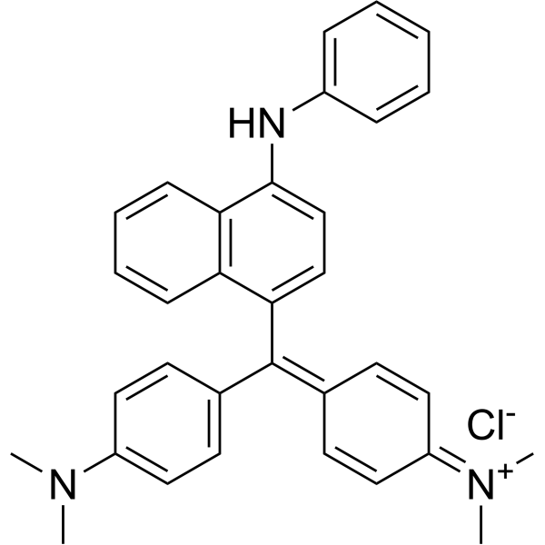
Basic blue 26 (Victoria blue B) is a synthetic cationic dye belonging to the class of triarylmethane dyes. It has a bright blue color and is commonly used as a colorant for a variety of applications, including textiles, paper and leather. Basic Blue 26 is also used as a biological stain for DNA and protein detection in laboratories. Due to its ability to bind negatively charged materials, it can be used as an indicator of the presence of specific molecules in biological samples. However, Basic blue 26 has been reported to have potentially harmful effects on human health and the environment and its use is regulated in some countries. Proper handling and disposal procedures are necessary to minimize its impact on the environment.
|
-
| Cat. No. |
Product Name |
Type |
-
- HY-D0914
-
|
FD&C Green No. 3; Food green 3; C.I. 42053
|
Dyes
|
|
Fast Green FCF is a sea green triarylmethane food dye, with absorption maximum ranging from 622 to 626 nm. Fast Green FCF is widely used as a staining agent like quantitative stain for histones at alkaline pH after acid extraction of DNA, and as a protein stain in electrophoresis. Fast Green FCF is carcinogenic and acts as a presynaptic locus by inhibiting the release of neurotransmitters in the nervous system .
|
-
- HY-W127770
-
|
|
Dyes
|
|
Pararosaniline hydrochloride is a pH-responsive basic dye, as a biological stain to track certain proteins. Pararosaniline hydrochloride has been used in the analysis of SO2 and formaldehyde and staining of bacteria or other organisms .
|
-
- HY-D0232
-
|
Brilliant Blue R
|
Dyes
|
|
Brilliant blue R250 (Brilliant Blue R), an anionic dye, is the most popular stain to detect proteins resolved in SDS-PAGE gels .
|
-
- HY-12489
-
|
Acid Red 112
|
Dyes
|
|
Ponceau S (Acid Red 112) is a non-specific protein dye commonly used as a stain for Western blot. Ponceau S is used in an acidic aqueous solution that is compatible with antibody-antigen binding and dyes the proteins on the membrane red .
|
-
- HY-D1544
-
|
|
Fluorescent Dyes/Probes
|
|
Uniblue A sodium is a reactive protein stain that can be used in the covalent pre-gel staining of the protein (Ex=594 nM) .
|
-
- HY-Y0700
-
|
|
Protein Labeling
|
|
Calconcarboxylic acid, an azo dye, acts as a silver-ion sensitizer to stain protein in SDS-PAGE gels. Calconcarboxylic acid increases silver binding on protein bands or spots by the formation of a silver-dye complex and also increases the reducing power o
|
-
- HY-D1475
-
|
|
Fluorescent Dyes/Probes
|
|
SIR-6-COOH is a fluorescent dye. SIR-6-COOH can be used for staining proteins in live-cell STED imaging
|
-
- HY-D1615
-
|
|
Dyes
|
|
BODIPY 530/550 NHS ester can be used for the stain of protein. BODIPY 530/550 NHS ester can be used for fluorescence OIM (oblique illumination microscopic) image .
|
-
- HY-D0987
-
|
|
Dyes
|
|
Stains-All, a cationic carbocyanine dye, is a convenient probe to study the structural features of the individual calcium-binding sites of calmodulin (CaM) and related calcium-binding proteins (CaBP) .
|
-
- HY-D1543
-
|
|
Fluorescent Dyes/Probes
|
|
Pyronin B is an organic cationic dye used for the staining of bacteria, mycobacteria and ribonucleic acids. Pyronin B is also used as a small hydrophobic (SH) protein channel inhibitor .
|
-
- HY-D1493
-
|
|
Fluorescent Dyes/Probes
|
|
FIM-1 is a fluorescent PKC (protein kinase C) probe that can be used for mitochondrial staining. FIM-1 inhibits PKC and acts as ATP-competitive catalytic site inhibitor .
|
-
- HY-D0166A
-
|
|
Dyes
|
|
Neutral Red (IND) is an organic dye commonly used in biology and cytology laboratories. It can be used to stain living cells, secreted proteins and other molecular structures, etc., and has a wide range of applications in cell imaging and staining. In addition, Neutral Red (IND) is widely used in industrial fields such as water treatment, food processing and paper manufacturing, for example as an indicator or colorant. Although the compound has no direct medical application, it has important application value in the fields of biology, chemistry and industry.
|
-
- HY-12489A
-
|
Acid Red 112, BS
|
Dyes
|
|
Both Ponceau S and Ponceau BS are synthetic dyes commonly used in biological research. They are commonly used as protein stains to visualize proteins in western blots and other protein detection analyses. Ponceau S is a red dye, while Ponceau BS is a blue shade of the same dye. Both dyes bind to positively charged amino acid residues in proteins for easy visualization. However, Ponceau S is more commonly used due to its higher sensitivity.
|
-
- HY-W127703
-
|
|
Fluorescent Dyes/Probes
|
|
Octadecyl Rhodamine B chloride is a cationic amphiphile that can be used for staining cell membranes. Octadecyl Rhodamine B chloride can be used in numerous studies including electronic energy transfer in organized molecular assemblies, membrane structure, and distances of closest approach between protein domains and membranes .
|
-
- HY-135056
-
|
|
Fluorescent Dyes/Probes
|
|
Mito-Tracker Green is a green fluorescent dye that selectively accumulates in the mitochondrial matrix. MitoTracker Green FM covalently binds mitochondrial proteins by reacting with free mercaptan of cysteine residues, allowing staining of mitochondrial membrane potential independent of membrane potential. Excitation/emission wavelength 490/523 nm.
|
-
- HY-W133919
-
|
|
Dyes
|
|
Aniline Blue sodium is a water-soluble dye commonly used as a biological stain for the detection of nucleic acids and proteins in various laboratory procedures such as electrophoresis and microscopy. Aniline Blue sodium has unique chemical properties that allow it to bind to specific cellular components, producing a color change that facilitates their visualization and analysis.
|
-
- HY-D0938
-
CFDA-SE
Maximum Cited Publications
53 Publications Verification
5(6)-Carboxyfluorescein diacetate succinimidyl ester; 5(6)-CFDA N-succinmidyl ester
|
Fluorescent Dyes/Probes
|
|
CFDA-SE is a fluorescent dye that can penetrate the cell membrane. It can react with the free amine group in the cytoskeleton protein inside the cell, and finally form a protein complex with fluorescence. After entering the cell, CFDA-SE locates in the cell membrane, cytoplasm and nucleus, and the fluorescence staining is strongest in the nucleus .
CFDA-SE dye can be uniformly inherited by the cells with cell division and proliferation, and its attenuation is proportional to the number of cell divisions. This phenomenon can be detected and analyzed by flow cytometry under the excitation light of 488 nm, and can be used to detect the proliferation of cells .
|
-
- HY-138658
-
|
JF526, SE; JF526, NHS
|
Fluorescent Dyes/Probes
|
|
Janelia Fluor 526, SE (JF526,SE) is a fluorogenic yellow fluorescent dye that contains NHS ester group. JF526 is a versatile scaffold for fluorogenic ligands, including labels for genetically encoded self-labeling protein tags and stains for endogenous structures . Janelia Fluor products are licensed under U.S. Pat. Nos. 9,933,417, 10,018,624 and 10,161,932 and other patents from Howard Hughes Medical Institute.
|
-
- HY-D0079
-
|
Hydroethidine; PD-MY 003
|
Fluorescent Dyes/Probes
|
|
Dihydroethidium, also known as DHE, is a peroxide indicator. Dihydroethidium penetrates cell membranes to form a fluorescent protein complex with blue fluoresces. After entering the cells, Dihydroethidium is mainly localized in the cell membrane, cytoplasm and nucleus, and the staining effect is the strongest in the nucleus. Dihydroethidium produces inherent blue fluorescence with a maximum excitation wavelength of 370 nm and a maximum emission wavelength of 420 nm; after dehydrogenation, Dihydroethidium combines with RNA or DNA to produce red fluorescence with a maximum excitation wavelength of 300 nm and a maximum emission wavelength of 610 nm. 535 nm can also be used as the excitation wavelength for actual observation .
|
-
- HY-D1804
-
|
|
Dyes
|
|
Vari Fluor 680-Streptavidin is a dye marker of Vari Fluor-streptavidin consisting of labeling streptavidin with a Vari Fluor series of fluorescent probes. Streptavidin is a high-affinity tetramer protein, each tetramer consisting of four identical streptavidin subunits. Streptavidin binds to biotin specifically via a reversible non-covalent effect. Streptavidin can achieve rapid and efficient detection of biotin markers, and is often used in immunofluorescence (IF), enzyme-linked immunosorbent assay (ELISA), immunohistochemical staining (IFH), in situ hybridization (ISH) and other experiments. Ex/Em=680 nm/701 nm.
|
-
- HY-D1805
-
|
|
Dyes
|
|
Vari Fluor 647-Streptavidin is a dye marker of Vari Fluor-streptavidin consisting of labeling streptavidin with a Vari Fluor series of fluorescent probes. Streptavidin is a high-affinity tetramer protein, each tetramer consisting of four identical streptavidin subunits. Streptavidin binds to biotin specifically via a reversible non-covalent effect. Streptavidin can achieve rapid and efficient detection of biotin markers, and is often used in immunofluorescence (IF), enzyme-linked immunosorbent assay (ELISA), immunohistochemical staining (IFH), in situ hybridization (ISH) and other experiments. Ex/Em=650 nm/665 nm.
|
-
- HY-D1806
-
|
|
Dyes
|
|
Vari Fluor 594-Streptavidin is a dye marker of Vari Fluor-streptavidin consisting of labeling streptavidin with a Vari Fluor series of fluorescent probes. Streptavidin is a high-affinity tetramer protein, each tetramer consisting of four identical streptavidin subunits. Streptavidin binds to biotin specifically via a reversible non-covalent effect. Streptavidin can achieve rapid and efficient detection of biotin markers, and is often used in immunofluorescence (IF), enzyme-linked immunosorbent assay (ELISA), immunohistochemical staining (IFH), in situ hybridization (ISH) and other experiments. Ex/Em=590 nm/617 nm.
|
-
- HY-D1807
-
|
|
Dyes
|
|
Vari Fluor 555-Streptavidin is a dye marker of Vari Fluor-streptavidin consisting of labeling streptavidin with a Vari Fluor series of fluorescent probes. Streptavidin is a high-affinity tetramer protein, each tetramer consisting of four identical streptavidin subunits. Streptavidin binds to biotin specifically via a reversible non-covalent effect. Streptavidin can achieve rapid and efficient detection of biotin markers, and is often used in immunofluorescence (IF), enzyme-linked immunosorbent assay (ELISA), immunohistochemical staining (IFH), in situ hybridization (ISH) and other experiments. Ex/Em=555 nm/565 nm.
|
-
- HY-D1808
-
|
|
Dyes
|
|
Vari Fluor 488-Streptavidin is a dye marker of Vari Fluor-streptavidin consisting of labeling streptavidin with a Vari Fluor series of fluorescent probes. Streptavidin is a high-affinity tetramer protein, each tetramer consisting of four identical streptavidin subunits. Streptavidin binds to biotin specifically via a reversible non-covalent effect. Streptavidin can achieve rapid and efficient detection of biotin markers, and is often used in immunofluorescence (IF), enzyme-linked immunosorbent assay (ELISA), immunohistochemical staining (IFH), in situ hybridization (ISH) and other experiments. Ex/Em=490 nm/515 nm.
|
-
- HY-D1809
-
|
|
Dyes
|
|
Vari Fluor 405-Streptavidin is a dye marker of Vari Fluor-streptavidin consisting of labeling streptavidin with a Vari Fluor series of fluorescent probes. Streptavidin is a high-affinity tetramer protein, each tetramer consisting of four identical streptavidin subunits. Streptavidin binds to biotin specifically via a reversible non-covalent effect. Streptavidin can achieve rapid and efficient detection of biotin markers, and is often used in immunofluorescence (IF), enzyme-linked immunosorbent assay (ELISA), immunohistochemical staining (IFH), in situ hybridization (ISH) and other experiments. Ex/Em=405 nm/431 nm.
|
-
- HY-W250147
-
|
Victoria blue B
|
Dyes
|
|
Basic blue 26 (Victoria blue B) is a synthetic cationic dye belonging to the class of triarylmethane dyes. It has a bright blue color and is commonly used as a colorant for a variety of applications, including textiles, paper and leather. Basic Blue 26 is also used as a biological stain for DNA and protein detection in laboratories. Due to its ability to bind negatively charged materials, it can be used as an indicator of the presence of specific molecules in biological samples. However, Basic blue 26 has been reported to have potentially harmful effects on human health and the environment and its use is regulated in some countries. Proper handling and disposal procedures are necessary to minimize its impact on the environment.
|
| Cat. No. |
Product Name |
Type |
-
- HY-D0232
-
|
Brilliant Blue R
|
Dyes
|
|
Brilliant blue R250 (Brilliant Blue R), an anionic dye, is the most popular stain to detect proteins resolved in SDS-PAGE gels .
|
-
- HY-D0014
-
|
|
Biochemical Assay Reagents
|
|
Brilliant Blue G-250 is a dye commonly used for the visualization of proteins separated by SDS-PAGE, offering a simple staining procedure and high quantitation. In the Bradford protein assay, protein concentrations are determined by the absorbance at 595 nm due to the binding of Brilliant Blue G-250 to proteins. Brilliant Blue G-250 is a safe highly selective P2×7R antagonist with promising consequent inactivation of NLRP3 inflammasome .
|
-
- HY-W011831
-
|
3-(Benzyldimethylammonio)propane-1-sulfonate; PDA
|
Surfactants
|
|
NDSB-256 is a non-stain remover sulfabetaine. NDSB-256 prevents protein aggregation and promotes denaturation of chemically and thermally denatured proteins .
|
Your information is safe with us. * Required Fields.
Inquiry Information
- Product Name:
- Cat. No.:
- Quantity:
- MCE Japan Authorized Agent:


